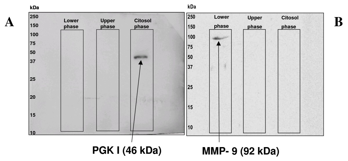
Figure 2

Western blot analysis for PGK-1 presence in the obtained fractions. Western blot analyses for the cytosolic enzyme phosphoglycerate kinase (PGK I), typical of the glycolysis, panel A, and for the membrane bound protein Metalloproteinase 9 (MMP9), panel B. The three fractions were run together on the same SDS-PAGE loading an equal amount of total protein. The same PVDF membrane was used. Film image with the relative protein bands only in the lane corresponding to the cytosolic compartment and to the membrane fraction are reported. Western blot images were captured by GS710 densitometer (Bio-Rad) and analyzed by QuantityOne software.
